Home
Uncategories
Foot Muscles Mri / Dorsal Muscles Of The Foot Anatomy And Function Kenhub / Muscle anatomy basics 12 photos of the muscle anatomy basics basics of muscle anatomy, muscle anatomy basics, human muscles, basics of muscle anatomy, muscle anatomy basics
Foot Muscles Mri / Dorsal Muscles Of The Foot Anatomy And Function Kenhub / Muscle anatomy basics 12 photos of the muscle anatomy basics basics of muscle anatomy, muscle anatomy basics, human muscles, basics of muscle anatomy, muscle anatomy basics
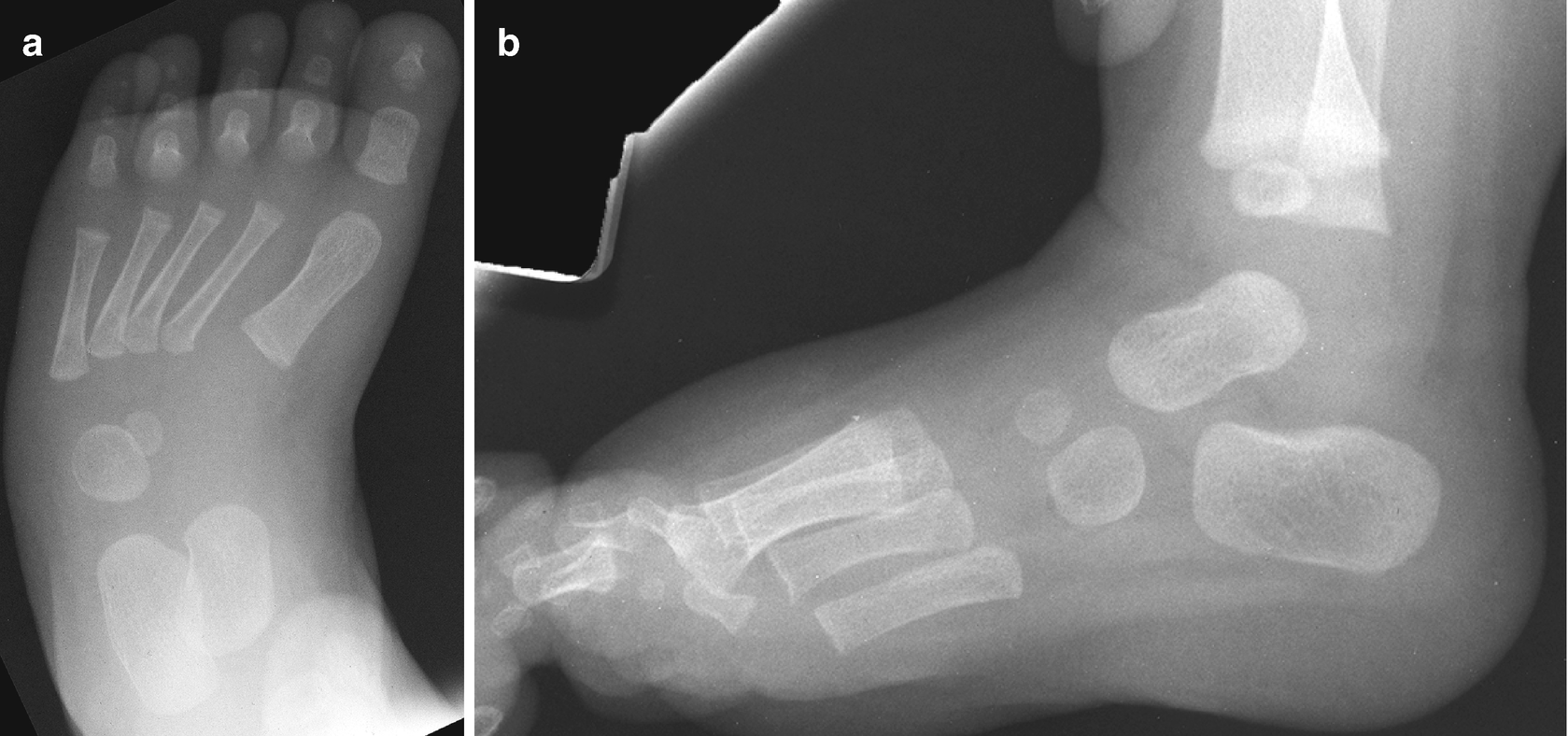
Foot Muscles Mri / Dorsal Muscles Of The Foot Anatomy And Function Kenhub / Muscle anatomy basics 12 photos of the muscle anatomy basics basics of muscle anatomy, muscle anatomy basics, human muscles, basics of muscle anatomy, muscle anatomy basics. One of the large muscles of the leg, it connects to the heel. In addition, an image of all the muscles of the back and plantar part of the foot, all tendons and tendon ligaments, blood vessels and nerves are obtained. They are mainly responsible for assisting some of the extrinsic muscles in their actions. It flexes and extends the foot, ankle, and knee. In the foot and ankle many accessory ossicles can be seen.
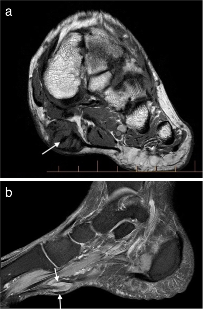
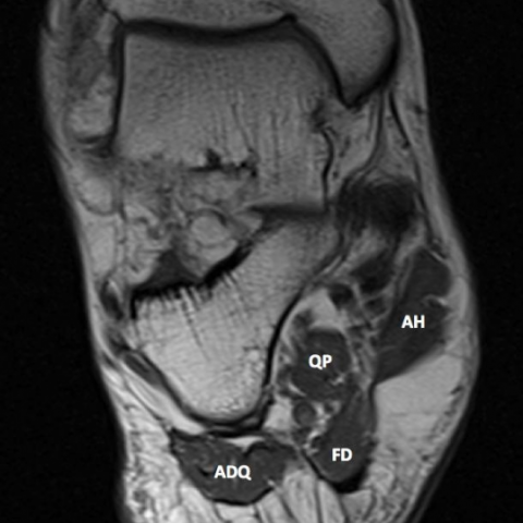
Those fibers of the most medial and largest belly are… The three plantar interossei muscles adduct the 3 rd, 4 th and 5 th toes toward the long axis through the 2 nd toe. Muscle injuries of the hip and thigh are a highly relevant issue in competitive sports imaging. Magnetic resonance imaging (mri) is the modality of choice in diagnosing accessory muscles, delineating their relationship to adjacent structures, and differentiating them from soft tissue tumors. Adductor hallucis is anatomically located in the central compartment of foot, but the muscle is functionally grouped with the medial plantar muscles of foot because it acts on the great toe (hallux).
Radiology Of The Pediatric Foot And Ankle Springerlink from media.springernature.com The presence of intramuscular edema (increased high t2/stir signal) on mri carries an extremely broad differential. This imaging technique assesses the ligaments and tendons, neurovascular structures (tarsal tunnel and plantar fascia), and the osseous structures(19). Radiologists need to be familiar with typical mri findings in order to accurately detect and classify muscle injuries. Mri is the choice of modality for further imaging the ankle and foot after obtaining initial radiographs. The studies were performed on a variety of magnets ranging from 0.2 to 1.5 t between march 15 and july 22, 2006. This small, thin muscle is absent in about. The machine uses radio waves and a magnetic field to generate images of the inside of the extremity in order to diagnose problems with the muscles, bones, joints, nerves, or blood vessels. Plantar plate of the foot:
Magnetic resonance imaging, otherwise known as mri, uses a combination of magnetic fields and radio waves to take images of the internal structures of your body.
Mri findings of acute turf toe: Denervation changes in muscles early. Both muscles are innervated by the deep fibular nerve. Proper interpretation of the findings is crucial, especially in elite athletes. The three plantar interossei muscles adduct the 3 rd, 4 th and 5 th toes toward the long axis through the 2 nd toe. Accessory muscles are isointense to skeletal muscle on all pulse sequences, and can insert by fleshy muscular or tendinous insertions. Anatomical structures of the ankle and foot and specific regions (major joints) are visible as dynamic labeled images. The intrinsic muscles of the foot are key contributors to foot function and are important to evaluate in lower limb disorders. Ultrasonography (us) affords high spatial resolution of muscle but is less sensitive than magnetic resonance (mr) imaging for mild edema and early myopathy. A case report and review of anatomy. Radiologists need to be familiar with typical mri findings in order to accurately detect and classify muscle injuries. This small, thin muscle is absent in about. Magnetic resonance imaging, otherwise known as mri, uses a combination of magnetic fields and radio waves to take images of the internal structures of your body.
A case report and review of anatomy. Those fibers of the most medial and largest belly are… Trauma effects of direct injury or tear denervation injury: Your doctor, with the help of a radiologist, can then examine these images to determine whether there is anything wrong with your foot or ankle. They are mainly responsible for assisting some of the extrinsic muscles in their actions.
Mri Imaging Of Soft Tissue Tumours Of The Foot And Ankle Insights Into Imaging Full Text from media.springernature.com With a muscle injury, for example, mri images often show a bright signal indicating that there is more water in the muscle, which is a sign of injury. Related posts of foot muscle anatomy mri muscle anatomy basics. An extremity mri is a type of scan used specifically for diagnostic imaging of the arm, leg, hand, or foot. Muscle anatomy basics 12 photos of the muscle anatomy basics basics of muscle anatomy, muscle anatomy basics, human muscles, basics of muscle anatomy, muscle anatomy basics The adductor hallucis has two heads: The gold standard in diagnostic imaging of muscle injuries is magnetic resonance imaging (mri). Your doctor, with the help of a radiologist, can then examine these images to determine whether there is anything wrong with your foot or ankle. Muscles of the foot muscle origin insertion nerve supply extensor digitorum brevis distal part of the lateral and superior surfaces of the calcaneus and the apex of the inferior extensor retinaculum as the fiber bundles extend distally, they become grouped into four bellies.
Related posts of foot muscle anatomy mri muscle anatomy basics.
This imaging technique assesses the ligaments and tendons, neurovascular structures (tarsal tunnel and plantar fascia), and the osseous structures(19). Mri findings of acute turf toe: Flexor digitorum brevis is in charge of the toe flexion at the metatarsophalangeal joints of the lateral four digits. The machine uses radio waves and a magnetic field to generate images of the inside of the extremity in order to diagnose problems with the muscles, bones, joints, nerves, or blood vessels. Routine ankle magnetic resonance imaging (mri) tests involve taking images of the foot and ankle in the axial, coronal, and sagittal planes parallel to the tabletop(2). Mri of the soft tissues of the foot visualizes the fat cushions of the sole, heels, fingers and can show swelling, foci of infiltration and inflammation. An extremity mri is a type of scan used specifically for diagnostic imaging of the arm, leg, hand, or foot. The three plantar interossei muscles adduct the 3 rd, 4 th and 5 th toes toward the long axis through the 2 nd toe. Findings on conventional arthrography and mr imaging. The presence of intramuscular edema (increased high t2/stir signal) on mri carries an extremely broad differential. Those fibers of the most medial and largest belly are… Muscle anatomy basics 12 photos of the muscle anatomy basics basics of muscle anatomy, muscle anatomy basics, human muscles, basics of muscle anatomy, muscle anatomy basics The studies were performed on a variety of magnets ranging from 0.2 to 1.5 t between march 15 and july 22, 2006.
While the total volume of plantar intrinsic foot muscles was similar in healthy and plantar fasciitis feet, atrophy of the forefoot plantar intrinsic foot muscles may contribute to plantar fasciitis by destabilizing the medial longitudinal arch. Ultrasonography (us) affords high spatial resolution of muscle but is less sensitive than magnetic resonance (mr) imaging for mild edema and early myopathy. Adductor hallucis is anatomically located in the central compartment of foot, but the muscle is functionally grouped with the medial plantar muscles of foot because it acts on the great toe (hallux). The majority of soft tissue lesions in the foot and ankle are benign. Those fibers of the most medial and largest belly are…
Baxter 039 S Neuropathy Isolated Fatty Atrophy Of The Abductor Digiti Minimi Muscle In Association With Plantar Fasciitis Eurorad from www.eurorad.org Proper interpretation of the findings is crucial, especially in elite athletes. The presence of intramuscular edema (increased high t2/stir signal) on mri carries an extremely broad differential. An extremity mri is a type of scan used specifically for diagnostic imaging of the arm, leg, hand, or foot. • muscle edema is seen secondary to multiple etiologies including trauma, infectious and inflammatory processes, autoimmune disorders, neoplasms, and denervation injuries • on mri muscle edema is characterized by increase in free water within the muscle • muscle edema is seen on mri as increased signal on fluid sensitive sequences t2 fs Plantar interossei (foot) dr yuranga weerakkody ◉ and dr geon oh et al. In the foot and ankle many accessory ossicles can be seen. 9 yao l, do hm, cracchiolo a, et al. The adductor hallucis has two heads:
Those fibers of the most medial and largest belly are…
Mri of the soft tissues of the foot visualizes the fat cushions of the sole, heels, fingers and can show swelling, foci of infiltration and inflammation. The studies were performed on a variety of magnets ranging from 0.2 to 1.5 t between march 15 and july 22, 2006. Radiologists need to be familiar with typical mri findings in order to accurately detect and classify muscle injuries. They are mainly responsible for assisting some of the extrinsic muscles in their actions. An extremity mri is a type of scan used specifically for diagnostic imaging of the arm, leg, hand, or foot. A case report and review of anatomy. Magnetic resonance imaging (mri) is the modality of choice in diagnosing accessory muscles, delineating their relationship to adjacent structures, and differentiating them from soft tissue tumors. It is classified to the central plantar muscles of the foot, together with quadratus plantae, lumbricals, plantar interossei and dorsal interossei muscles. Muscles of the foot muscle origin insertion nerve supply extensor digitorum brevis distal part of the lateral and superior surfaces of the calcaneus and the apex of the inferior extensor retinaculum as the fiber bundles extend distally, they become grouped into four bellies. Mri findings of acute turf toe: This small, thin muscle is absent in about. This imaging technique assesses the ligaments and tendons, neurovascular structures (tarsal tunnel and plantar fascia), and the osseous structures(19). The majority of soft tissue lesions in the foot and ankle are benign.
0 Comments:
Posting Komentar